Cuddly cats, toxoplasma and pregnancy

DCF 1.0
DCF 1.0

A recent case of severe toxoplasma infection in pregnancy at one of my institutions reinforces the need for preconception and early pregnancy hygiene advice.

What is toxoplasma infection?

Toxoplasmosis is an infection caused by the parasite Toxoplasma gondii. The parasite may be found in raw and cured meat, raw eggs, unpasteurised milk and cat faeces. It can also be found in soil contaminated with cat faeces. This means cat owners need to be careful when they handle kitty litter and should wear gloves when they garden in areas where the soil may be contaminated.

Clinical consequences of toxoplasma infection

Outside of pregnancy, toxoplasmosis results in a mild flu-like illness or a subclinical infection with no symptoms.

However, toxoplasma infection acquired in the period from three months prior to conception and during pregnancy, is more significant.  The parasite can cross the placenta and infect the developing baby who has an immature immune system. The consequences can be serious and include miscarriage, brain damage, blindness and death.

Why the timing of the infection is important

The timing of the infection in pregnancy influences the risk of the baby acquiring the infection. It also impacts on the severity of the sequelae of the infection in the baby.

In early pregnancy, less than 5% of babies are infected. This is because the placenta is too immature for transplacental infection.

However, in late pregnancy, 65% of babies can acquire the infection, as the placenta is mature and rates of transplacental infection are higher.

Even though infection caught late in pregnancy is more likely to result in fetal infection, the consequences of the infection are usually less severe.

In contrast,  infection caught by the mother early in her pregnancy results in a lower fetal infection rate but has far more significant consequences for her baby.

Most of the advice on toxoplasma in pregnancy comes from a few landmark research papers. It is worth looking at the findings from these papers to help guide pregnancy advice and management.


Landmark research papers

1. Pregnancy outcomes

The first landmark paper on toxoplasma in pregnancy was published in the New England Journal of Medicine in 1974 (1).

The researchers reported on the pregnancy outcomes of two groups of women. One group had evidence of toxoplasmosis prior to pregnancy and the other group acquired toxoplasmosis during pregnancy.

The key findings were:

  • Mothers who acquired toxoplasmosis prior to pregnancy did not infect their baby. No babies in this group had toxoplasmosis.

However;

  • In the 77 women who acquired toxoplasma infection during pregnancy:
    • 11 women miscarried;
    • 7 women suffered fetal death in utero and delivered a stillborn baby;
    • 2 newborns died soon after delivery;
    • 7 babies had severe effects of congenital toxoplasmosis and had cerebral and ocular complications;
    • 11 babies had  only mild illness requiring treatment:
    • 39 babies had subclinical illness with no long term problems.
  • Severe outcomes were only detected when the maternal toxoplasma infection occured during the first two trimesters (up to 27 weeks pregnancy).
  • Acquiring the toxoplasma infection in the third trimester resulted in subclinical infection or in no fetal infection.

2. Risk factors

The largest published research paper on risk factors comes from France (2), which has one of the highest rates of toxoplasma infection in pregnancy in the world. The authors reported that there were approximately 4900 cases of primary Toxoplasma infection in pregnant women in France each year.

Since 1992, France has had a policy that all pregnant women at risk of Toxoplasma infection undergo monthly serological testing. This research paper reported on 80 pregnant women who seroconverted to Toxoplasma in pregnancy and compared their risk factors against 80 pregnant women who had repeatedly negative tests.

After performing a multivariate statistical analysis to ensure differences between the two groups of women were accounted for, they found the risk factors for Toxoplasma infection were:

  • Poor hand hygiene increased risk by almost 10 fold (OR=9.9; 95%CI: 0.8-125);
  • Consumption of undercooked beef increased risk by almost 6 fold (OR=5.5; 95%CI: 1.1-27);
  • Having a pet cat increased risk by almost 5 fold (OR=4.5; 95%CI: 1.0-19.9);
  • Frequent consumption of raw vegetables outside the home increased risk by 3 fold (OR=3.1; 95%CI: 1.2-7.7);
  • Consumption of undercooked lamb increased risk by 3 fold (OR=3.1; 95%CI: 0.85-14).

The researchers noted that when women were provided with documentary advice on how to prevent toxoplasma infection, they had a lower risk of infection. They concluded that all  pregnant women should be given information on their eating habits, hand hygiene and cats in order to reduce the risk of acquiring toxoplasma infection in pregnancy.

3. Treatment of toxoplasma infection in pregnancy

The third landmark paper addressed the treatment of toxoplasma infection in pregnancy (3).

This treatment study was conducted across 5 major centres that specialised in the management of toxoplasma infection in pregnancy. The aim of the study was to determine if prenatal antibiotic therapy could reduce the rate of mother to baby transmission of Toxoplasma gondii and reduce the risk of severe consequences in infected babies as measured when they were one year old.

The study followed 144 women and considered key factors such as the gestational age at which the infection was acquired, the administration of antibiotic therapy, duration of antibiotic therapy, and time lapse between infection and the start of antibiotic therapy.

The authors reported that 64 of the 144 infected women (44%) gave birth to a congenitally infected infant.

After performing a multivariate analysis they concluded that the rate of transmission from the mother to the baby was not affected by the administration of antibiotics. The only factor that affected the rate of transmission was the gestational age at which the mother developed the infection in pregnancy (P < .0001).

However, antibiotic administration significantly reduced the risk of serious sequelae in the babies (P = 0.026, odds ratio 0.30, 95% confidence interval 0.104-0.863).

Of particular note, severe sequelae were significantly reduced when the mother was administered antibiotics (P = .007, odds ratio 0.14, 95% confidence interval 0.036-0.584). The sooner antibiotics were given after the infection, the less frequently sequelae were seen (P = 0.021).

The results were consistent with the earlier New England Journal of Medicine paper which also reported that treatment of infected mothers with the antibiotic Spiramycin reduced the impact of fetal infection (1).

What is the current pregnancy advice for preventing toxoplasmosis in pregnancy?

In Australia, the risk of toxoplasma infection is low. Therefore routine screening for the infection is not performed.

The main method to prevent infection is through education on strategies to reduce the risk of becoming infected.

The Australian Government’s Pregnancy, Birth and Baby website outlines strategies to reduce the risk of infection (4). These are:

  • Wear gloves when gardening, particularly when handling soil;
  • Wash your hands thoroughly after handling soil, using soap and hot water;
  • Do not eat raw or undercooked meat;
  • Cook all red meat until no trace of pinkness remains and the juices run clear;
  • Do not eat cured meats (e.g. Parma ham, salami);
  • Do not taste meat before it is fully cooked;
  • Wash your hands thoroughly after handling raw meat;
  • Wash all kitchenware thoroughly after preparing raw meat;
  • Always wash fruit and vegetables before cooking and eating;
  • Avoid drinking unpasteurised goat’s milk or eating products that are made from it;
  • Do not handle or adopt stray cats;
  • Keep animals, especially cats, away from areas that you prepare/store food;
  • Wash your hands and other exposed body parts with soap and running water after touching animals, their enclosures or food containers;
  • Avoid cat faeces in cat litter or soil – wear gloves if you are changing a cat’s litter tray and wash your hands thoroughly afterwards. If you are pregnant or immune deficient, ask someone else to change it for you and empty the litter tray daily;
  • Feed your cat dried or canned cat food rather than raw meat;
  • Cover children’s outdoor sand boxes to prevent cats using them as litter boxes;
  • Avoid contact with sheep and newborn lambs during the lambing season.

Management if infection is suspected

If a pregnant woman is concerned that she may have acquired toxoplasmosis in pregnancy, she should immediately see her doctor or midwife to discuss her concerns and be referred for an antibody test.

If the test demonstrates the presence of IgG antibodies, she can usually be reassured that she has already had the infection and is immune.

If the test demonstrates IgM antibodies, or no antibodies but the clinical suspicion of infection is high, the pregnancy should be managed by an obstetrician with expertise in this area.

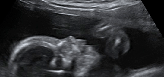
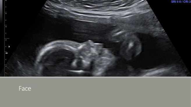
Many cases are not detected until an ultrasound examination is performed and is noted to be abnormal.

Once maternal infection is diagnosed, or an ultrasound examination is suspicious of features of toxoplasma infection in the fetus, a tertiary level ultrasound, blood tests and amniocentesis to diagnose fetal infection are indicated. Antibiotic therapy needs to be commenced as soon as possible to reduce the impact of the fetal infection.

All cases should be managed in a tertiary obstetric hospital and advice and management will be individualised to each woman.

Babies should be enrolled in a follow up service to ensure any sequelae are diagnosed and managed as soon as possible.

Summary

Toxoplasma infection in pregnancy resulting in severe consequences for the baby is rare. However it can be prevented with good hygiene practices and managed with expert interventions by specialist teams.

The most important message is to be aware of good hand hygiene practices.

References

  1. Dismounts G, Couvreur J. Congenital Toxoplasmosis — A Prospective Study of 378 Pregnancies.N Engl J Med 1974; 290:1110-1116May 16, 1974. DOI: 10.1056/NEJM197405162902003
  2. Baril L, Ancelle T, Goulet V, Thulliez P, Tirard-Fleury V, Carme B. Risk Factors for Toxoplasma Infection in Pregnancy: A Case-Control Study in France. 
    Scandinavian Journal of Infectious Diseases 1999; 31(3): 305-309. 
    doi.org/10.1080/00365549950163626

When having a baby can kill you

Slide10

Pregnancy is usually a happy time with the outcome being a healthy baby. However, some complications in pregnancy can be serious. Over the years I have cared for many couples with molar pregnancy (technically known as gestational trophoblast disease). This terrible complication of pregnancy not only results in grief from the “lost baby”, but can also have lasting physical, social and psychological consequences for both the mother and father (1,2,3,4). Untreated, molar pregnancy can cause death of the mother (1,2).

Of note, our research into molar pregnancy revealed that many fathers experienced lasting social and psychological symptoms following molar pregnancy (4). One reason for this is due to the origins of the disease.

Origins of molar pregnancy

There are two types of molar pregnancy, a complete mole and a partial mole. In both cases, the male sperm plays a key role.

In a complete molar pregnancy, sperm (one or two) fertilize an egg that has lost its female genetic material (DNA). Therefore all the genetic material in the fertilized egg arises from the male and none from the female.

In a partial molar pregnancy, a single egg is fertilized by two sperm causing an excess of male genetic material within the fertilised egg.

The incidence of molar pregnancy varies around the globe, from 1 in 200 to 1 in 2000 pregnancies (1,2).

Impact on pregnancy

Sadly, molar pregnancy never results in a normal baby except for the extraordinarily rare cases of twinning where one twin is a molar pregnancy and the other a normal pregnancy. Instead, the usual situation is that the uterus becomes full of abnormal placental tissue and no baby is present (complete mole) or some fetal development occurs, but the fetus  is  malformed and not viable (partial mole).

The abnormal placental tissue causes bleeding and can metastasis around to body to other organs such as the lungs, in the same way an untreated cancer may spread around the body.

Impact on women

Most molar pregnancies present with abnormal vaginal bleeding between 8 and 16 weeks of pregnancy. Initially most women are concerned about miscarriage.

The diagnosis may be strongly suspected following an ultrasound, where a characteristic pattern called a “snowstorm” may be seen within the uterus. However, the condition is not definitively diagnosed until a sample of the tissue within the uterus is sent for analysis (histopathology) and tissue that looks like a cluster of grapes (abnormal chorionic villi) are seen under the microscope.

Some women may present with signs of thyroid disease, as the abnormal placental tissue can produce thyroid-like hormones. Women may also present with excessive nausea and vomiting of pregnancy (hyperemesis) and rarely may present with abnormally high blood pressure readings under 20 weeks of pregnancy.

Management of molar pregnancy

Once a molar pregnancy is confirmed, management involves surgery, follow up surveillance and possibly chemotherapy.

The initial management is uterine suction curettage. This surgical procedure is necessary to confirm the diagnosis and exclude an even rarer form of gestational trophoblastic disease called choriocarcinoma. The surgery carries more risk than a usual suction curettage, as the abnormal placental tissue is very vascular, and therefore the risk of heavy bleeding is higher. This means that the attending gynaecologist will often cross match blood and organise an anaesthetic consultation to plan the safest time to perform surgery. Medication may be required following surgery to help contract the uterus and reduce post operative bleeding.

Following surgery, women receive a “risk rating” that is determined on a number of factors such as their levels of pregnancy hormone, blood group, the presence of metastatic disease and the histopathology of the molar pregnancy.

Based on the “risk rating” results, women enter a follow up surveillance program that involves monitoring with serial blood or urine pregnancy hormone levels.

If a women had a high initial risk score, or her pregnancy hormone levels rose or failed to fall during her surveillance period, then she will require chemotherapy. This is usually Methotrexate, but in some case will be combined chemotherapy.

The impact of molar pregnancy on women is often profound. This is particularly true as the risk of molar pregnancy increases as women become older. Some women may have been trying to conceive for many years and then discover their pregnancy is a molar pregnancy. Not only do they not have the baby they desire, they face surgery, prolonged surveillance during which pregnancy is contraindicated, and possible chemotherapy. They must defer trying to have a child until they have been cleared (3,4).

Impact on partners

Partners of affected women can also suffer due to delayed childbearing, prolonged stress and a feeling of guilt related to the male role in the origins of molar pregnancy (4,5,6).

In our research, we contacted 158 former patients in our service with molar pregnancy and through these women, interviewed  41 partners. We found many partners were as emotionally fragile as the woman. For full results click here.

In a thematic analysis we found several themes related to anxiety and fear, sadness and depression, and guilt.

Anxiety

Anxiety was the dominant theme, rather than depression. Anxiety arose in male partners from a sense of frustration consequent to experiencing loss of control over their fertility, particularly their anxiety that they, as a couple, may never have a child.

‘Wouldn’t have occurred to us before when we were just worried about possible health of a baby’

‘Words cannot describe how emotionally stressful it was… I witnessed my partner being torn apart emotionally.’

‘Almost given up hope/plans of having a child at our age (maybe still some fear that another pregnancycould go awry).’

‘My world came crashing down.’

Guilt and blame

Partners felt guilty or blamed themselves for the occurrence of the molar pregnancy. Factors such as the male contribution in conception and individual genetic structures impacted on male participant’s view of cause and effect.

‘I somehow feel responsible in a way that it may have been my fault that it had something to do with my (works) my body that wasn’t right that caused this unusual pregnancy.’

Medical care

Themes relating to medical care centred around the actual treatment of molar pregnancy and the constant reminder of the diagnosis during the prolonged follow up perios that meant couples relived the experience. This delayed emotional recovery. The lack of clear information added to confusion and uncertainty.

‘…we are constantly reminded of our ‘failure’ through monthly urine samples, etc.’

‘I didn’t know as a individual at the time what was going on with my partner because we didn’t have enough information.’

Male partners’ displacement of feelings

Male partners felt a sense of indirect involvement in the management of the molar pregnancy. A new unfamiliar distancing occurred in a small percentage of couple relationships because of withdrawal from communication with partners during this time. This left partners feeling hopeless, unable to initiate appropriate actions to help their partner cope with trauma resulting from the diagnosis.

Partners felt they had to manage other financial and social matters additionally during this period of time. The male partner viewed himself largely as a supporter and made a distinction from being the patient.

‘I still cannot imagine what it would be like for my partner as she was the one carrying the pregnancy.’

‘It is hard for the husband to feel the same sense of loss as the wife because he has not had any physical contact with the “baby”.’

‘I felt very detached from it because it wasn’t my body going through the miscarry.’

Sexual function

Some men reported disparity in sexual functioning with their partner and described sexual tensions in their relationship.

‘My partner seems a little numb now, compared with before, and that makes it harder to feel good about sex and being close. I’m still keen but she seems less so…’

‘With all this happening inside her, she now seems less interested in sex, maybe that’s normal, but when I try she looks almost scared.’

‘…reduced desire by wife/apprehension re. sexual, even sensual contact…’

Positive role of children

The protective effect of children came through in our research. Subsequent delivery of a healthy child overcame the sense of loss.  This was reported both as an actual experience and as a hypothetically positive experience.

‘The scars only really started healing once we were given the gift of a beautiful baby boy nearly 2 years later.’

Summary

Most of the time pregnancy is a happy event, but occasionally things go wrong. It is important to remember that both mother and father may be deeply impacted and to provide support and follow up when things don’t go to plan.

Ultimately, providing support to ensure the couple are able to help each other through a sad and frightening experience is as important as getting the actual medicine right.

 References

  1. Berkowitz RS, Goldstein DP. Gestational trophoblastic diseases. In Principles and Practice of Gynecologic Oncology, Hoskins WJ, Perez CA, Young RC (eds.), Lippincott Williams & Wilkins: Philadelphia, PA, 2000; pp 1117–1137.
  2. Feltman CM, Growden WB, Wolfberg AJ et al. Clinical characteristics of persistent gestational trophoblastic disease after partial hydatidiform molar pregnancy. J Reprod Med 2006;51:902–906.
  3. Berkowitz RS, Marean AR, Hamilton N et al. Psychological and social impact of gestational trophoblastic neoplasia. J Reprod Med 1980;25:14–16.
  4. Quinlivan JA, Ung KA, Petersen RW. The impact of molar pregnancy on the male partner.Psychooncology. 2012 Sep;21(9):970-6. doi: 10.1002/pon.1992. Epub 2011 May 24.
  5. Wenzel L, Berkowitz RS, Robinson S, Bernstein M, Goldstein D. The psychological, social, and sexual consequences of gestational trophoblastic disease. Gynecol Oncol 1992;46:74–81.
  6. Wenzel L, Berkowitz RS, Robinson S, Goldstein DP, Bernstein MR. Psychological, social and sexual effects of gestational trophoblastic disease on patients and their partners. J Reprod Med 1994;39(3):163–167.

Pokemon Go, exercise and gestational diabetes mellitus

IMG_0675

The recent Pokemon Go craze could have an unintentional benefit for women with pregnancy complicated by Gestational Diabetes Mellitus (GDM). The exercise involved in walking  around parks trying to capture Pokemon helps manage blood sugar levels and can lead to a reduced need for medication and diabetic complications.

The benefits of exercise in pregnancy

Regular exercise, particularly walking, is beneficial in pregnancy. Not only can regular exercise limit excessive gestational weight gain to international standards, it can also help prevent or manage GDM (1,2).

However, encouraging women to participate in regular exercise during pregnancy has proven challenging (3). Several randomised trials of exercise interventions in pregnancy have failed to demonstrate an effect on preventing excessive gestational weight gain or on the incidence or management of GDM, mainly due to poor compliance and low levels of participation by pregnant women in exercise programs (3,4,5).

So how might Pokemon Go help?

Women diagnosed with GDM are often asked to monitor their blood sugar level each morning (fasting blood sugar level) and again  2 hours after every meal (post prandial 2 hour blood sugar level). If the morning fasting blood sugar level, or the post prandial 2 hour blood sugar levels are higher than recommended targets (2,5), then medication may be necessary in order to reduce the risk of pregnancy complications such as abnormal fetal growth (macrosomia), excessive amniotic fluid (polyhydramnios), placental damage or fetal death in utero.

Regular exercise, even a 30 minute walk performed three times a week, can be helpful in regulating gestational weight gain and blood sugar levels.

This is where Pokemon Go might help.

By combining a regular walk with a game, it might encourage pregnant women to walk.

Maybe the manufacturers could invent pregnant Pokemon for our pregnant women to capture and provide an extra incentive!

Over to you game makers.

What else is new in gestational diabetes research?

Our research team recently published a paper on managing gestational diabetes (6). In this research study we explored whether it was possible to safely streamline the number of women who have to undergo antenatal investigations.

The particular focus of our recent study was on the value of fetal cardiotocography (CTGs) in managing GDM (6).

Different levels of risk in GDM?

The prevalence of GDM is rising due to increases in maternal obesity and a rise in sedentary lifestyles (6,7,8). If increasing numbers of pregnant women need increasing numbers of tests, our maternity systems will explode and costs of care will rise.

GDM pregnancies do have an increased risk of maternal and fetal complications such as gestational hypertension, pre-eclampsia, caesarean delivery, development of type 2 diabetes postpartum, fetal macrosomia, birth trauma and shoulder dystocia (6,7,8). The risk of maternal and fetal complications is particularly high in GDM pregnancies with poor blood sugar control (6,7,8).

Medications that reduce blood sugar levels in women with GDM include Insulin and Metformin. Medication is only prescribed when women cannot achieve ideal blood fasting and 2-hour post prandial blood sugar levels despite eating a diabetic diet and undertaking regular exercise.

Women who need medication to manage their blood sugar level are therefore  at higher risk of potential pregnancy complications compared to women who are able to manage their blood sugar levels with  diet and exercise.

Fortunately, 70% of women can manage their blood sugar levels with diet and exercise. This means only 30% of women diagnosed with GDM require medication.

Antenatal monitoring in GDM

Antenatal fetal monitoring is routinely performed in pregnancies complicated by GDM.

The two most common tests undertaken to monitor the wellbeing of the fetus are cardiotocographs (CTG) and ultrasound (9,10).

CTG can detect some pregnancies at risk of stillbirth, allowing for prompt further intervention (9,10).

How do CTGs work?

The heart rate of a fetus is determined by a balance between two different types of neurotransmitters (sympathetic and parasympathetic) that  act on the sinoatrial node in the heart (11).

This balance is mediated through a number of factors including catecholamines (11). If fetal pathology is present, and the fetus is unwell, this balance can be affected, and changes in fetal heart rate patterns can be observed on a heart rate trace – the CTG (11,12). 

A number of conditions are associated with abnormal CTG tracings. Specific CTG findings that suggest fetal hypoxia and acidosis include reduced variation in the baseline fetal heart rate and loss of rises in heart rate (accelerations) or development of drops in fetal heart rate after uterine contractions (late decelerations) (13).

Using CTGs in pregnancy complicated with GDM

As high levels of blood sugar can damage the placenta and lead to fetal pathology that makes the fetus at risk of low oxygen or death, CTGs have been used to monitor GDM pregnancies.

However, there is a lack of consensus on the frequency and commencement gestation of CTG monitoring in GDM pregnancies (14,15,16).

Results from our research

In our recent research publication, published in the Australian and New Zealand Journal of Obstetrics and Gynaecology, we evaluated the role of CTGs in managing pregnancy complicated by GDM (6). Click here to read to full paper.

We audited 1404 consecutive antenatal CTGs in women diagnosed with GDM to determine how often they resulted in a change in management.

Overall, we found that in women requiring medication in order to manage their blood sugar levels, 43 CTGs were required to change management.

In women who did not require medication to manage their blood sugar levels, but who had another factor complicating their pregnancy, 161 CTGs were required to change management.

However, in women who did not require medication to manage their blood sugar levels and who had no other pregnancy complication, CTGs did not change management.

Therefore, if pregnant women with GDM can achieve good blood sugar control with changes to their diet and exercise, they do not require CTG monitoring.

This further emphasises the need to promote a diabetic diet and regular exercise in women with GDM.

If Pokemon Go is a potential solution to help encourage pregnant women walk and exercise on a regular basis, then it might result in a cost saving to our health system through improved pregnancy outcomes, less need for prescribed medication in GDM pregnancy and less need for antenatal monitoring with CTGs.

Maybe we should run a trial?

References

  1. Quinlivan J. The Challenge to deliver cost effective care for patients with Gestational Diabetes Mellitus. Repro Syst Sexual Dis 2014; 2014(3):4. DOI: 10.4172/2161-038X.1000144
  2. International Association of Diabetes and Pregnancy Study Groups Recommendations on the Diagnosis and Classification of Hyperglycemia in pregnancy. Diabetes Care 2010;33:676-682.
  3. Quinlivan J Dietary component of lifestyle interventions helps obese pregnant women. Evidence Based Medicine 06/2012; 18:e4 doi:10.1136/eb-2012-100794.
  4. Quinlivan J, Juliania S, Lam L. Antenatal dietary interventions in obese pregnant women to restrict gestational weight gain to institute of medicine recommendations: a meta-analysis. Obstetrics and Gynecology 2011: 118(6): 1395-401.
  5. Nankervis A, McIntyre HD, Moses R, Ross GP, Callaway L, Porter C, et al. Consensus guidelines for the testing and diagnosis of gestational diabetes mellitus in Australia. Australasian Diabetes in Pregnancy Society; 2014.
  6. Jeffery T, Petersen RW, Quinlivan JA. Does cardiotocography have a role in the antenatal management of pregnancy complicated by gestational diabetes mellitus? ANZJOG 2016; DOI: 10.1111/ajo.12487.
  7. Landon MB, Mele L, Spong CY, Ramin SM, Casey B, Wapner RJ, et al. The relationship between maternal glycemia and perinatal outcome. Obstet Gynecol. 2011 Feb;117(2):218-24.
  8. HAPO Study Cooperative Research Group. Hyperglycemia and adverse pregnancy outcomes. N Engl J Med. 2008;358:1991-2002.
  9. Graves CR. Antepartum fetal surveillance and timing of delivery in the pregnancy complicated by diabetes mellitus. Clin Obstet Gynecol. 2007;50(4): 1007-1013.
  10. Kjos SL, Leung A, Henry OA, Victor MR, Paul RH, Medearis AL. Antepartum surveillance in diabetic pregnancies: predictors of fetal distress in labor. Am J Obstet Gynecol. 1995 Nov;173(5):1532-1539.
  11. McDonnell S, Chandraharan E. The Pathophysiology of CTGs and Types of Intrapartum Hypoxia. Current Women’s Health Reviews. 2013;9(3): 158-68.
  12. McDonnell S, Chandraharan E. Fetal Heart Rate Interpretation in the Second Stage of Labour: Pearls and Pitfalls. Br J Med Med Res. 2015;7(12): 957-70.
  13. Devoe LD, Jones CR. Nonstress test: evidence-based use in high-risk pregnancy. Clin Obstet Gynecol. 2002 Dec;45(4):986-992.
  14. Metzger BE, Buchanan TA, Coustan DR, de Leiva A, Dunger DB, Hadden DR, et al. Summary and Recommendations of the Fifth International Workshop-Conference on Gestational Diabetes Mellitus. Diabetes Care. 2007 Jul;30(S2) :S251-60.
  15. Landon MB, Vickers S. Fetal surveillance in pregnancy complicated by diabetes mellitus: is it necessary?. J Matern Fetal Neonatal Med. 2002 Dec; 12(6):413-416.
  16. Loomis L, Lee J, Tweed E, Fashner J. What is appropriate fetal surveillance for women with diet-controlled gestational diabetes?. J Fam Pract. 2006 Mar;55(3):238-240.


Causes and consequences of too many antibiotics in pregnancy

Slide10

Antibiotics in pregnancy

I was recently checking audited hospital files from one of my research trials and noticed the majority of  women had been prescribed an antibiotic in pregnancy. The commonest indication was for prophylaxis against wound infection in women about to have a caesarean section. The second commonest indication was to prevent neonatal group B streptococcal infection in women with vaginal colonisation.

However, many other women had been prescribed antibiotics for urinary tract, vaginal and respiratory infections.

Yet the audit demonstrated that many of the mid stream urine samples and vaginal swabs collected from these women were ultimately normal. The respiratory infections documented in the notes were almost universally viral, and not bacterial.

It seems antibiotics in pregnancy are being overprescribed.

Overprescription of antibiotics in pregnancy

No drug or medication should be taken in pregnancy unless benefit outweighs the risk of harm.

A course of antibiotics prescribed for a viral chest infection or for urinary symptoms when no infection is present, is not harmless. Many patients and obstetric clinical staff mistakenly think it is “safer” to use antibiotics when in doubt.

However, there is a risk of real harm from the overprescription of antibiotics – harm to the mother, baby, and wider community.

Harm to the mother

Every time antibiotics are prescribed, they kill bacteria. However, they kill both harmful and beneficial bacteria.

In pregnancy, the flora of vagina stabilises (1). This stabilisation is associated with improved outcomes in pregnancy.

The use of antibiotics disrupts normal vaginal flora. This is because beneficial bacteria die and this creates an opportunity for pathogenic bacteria to colonise and infect the vagina.  The presence of pathogenic vaginal bacteria in pregnancy has  been linked to many adverse outcomes including preterm birth and fetal death in utero (2,3,4,5).

Colonisation by pathogenic bacteria is not the only risk. Other micro-organisms, such as fungi, can colonise or infect the vagina following a course of antibiotics. One commonly observed complication is post antibiotic vaginal Candidal infection. This can cause symptoms of itch, soreness and vaginal discharge.

Harm to the baby

As a baby is born, it passes through the vagina and acquires microorganisms from the mother’s reproductive tract. When the mother’s vaginal flora is healthy and stabilised, the baby’s microflora in the gastrointestinal tract will also be healthy and stabilise.

However, pathogenic bacteria in the maternal genital tract can be directly transmitted to the baby, causing serious newborn infection.

Even relatively minor pathogens such as Candida can still cause postnatal problems.  Vaginal Candidal infection can result in neonatal oral Candidal infection, and Candidal infection of the maternal nipples. The end result can be breastfeeding and overall feeding difficulties.

Harm to the community

The biggest harm arising from the overprescription of antibiotics comes from the development of resistance.

The Clinical Senate of Western Australia recently hosted a policy debate to develop recommendations to prevent the development and transmission of “Superbugs”. These are bacteria that are resistant to many antibiotics (6).

The World Health Organisation’s 2014 report on global surveillance of antimicrobial resistance reported that antibiotic resistance is no longer a prediction for the future; it is a reality. This year, several bacteria have been identified that are resistant to every known antibiotic.

This is a ticking time bomb for humanity.

Without urgent, coordinated action, the world is heading towards a post-antibiotic era in which common infections and minor injuries, which have been treatable for decades, can once again kill.

Impact of infections and antibiotic resistance

According to the National Health and Medical Research Council (NMHRC) and Australian Commission on Safety and Quality in Healthcare (NSQHS), infection is the most common complication affecting hospital patients, affecting 200,000 patients per year (7). At least half of healthcare associated infections are preventable. Successful infection control to minimise the risk of transmission requires a range of strategies across all levels of the healthcare system and a collaborative approach for successful implementation.

Excess length of stay due to a surgical site infection is between 3.5 and 23 hospital bed days, depending on the type of infection. The total national number of bed days due to surgical site infections for a one year period was estimated to be 206,527 bed days (8). If there was optimal use of antimicrobials and containment of antimicrobial resistance, $300 million of the Australian national healthcare budget could be redirected to more effective use every year (9).


What are Superbugs?

Dr Paul Armstrong, Director, Communicable Diseases Control Directorate, Public Health Division, Department of Health WA recently defined Superbugs as being multi-resistant organisms (MROs), resistant to a number of antibiotics. MROs arose from natural selection, that is, evolutionary pressure that selected resistant organisms following exposure to antibiotics within human medicine, veterinary medicine and agriculture.

Dr Armstrong stated the pressure on bacteria to develop resistance occurred in both hospitals (especially large tertiary hospitals) where the sickest patients are cared for and where the need for powerful antibiotics is greatest, and in the community.

Antibiotic resistance organisms are, to some extent, a natural process. However, overuse and misuse of antibiotics accelerates the emergence of drug-resistant strains, so that a drug that was previously effective to treat a particular microorganism is rendered ineffective.

Cost of Superbugs

Dr Armstrong told the Clinical Senate of Western Australia that the USA Centers for Disease Control and Prevention (CDC) estimated that $20 billion in direct costs was associated with antimicrobial resistance each year.

There is a direct cost on the health system due to increased costs of antibiotics, special equipment, prolonged length of stay, increased staff time and tying up of resources.

Some bacteria now had no antibiotics effective against them.

Origins of Superbugs

There are three major sources of Superbugs.

The first is  environmental contamination with antibiotics. This is is a particular problem in developing countries that manufacture pharmaceuticals and where the use of antibiotics in agriculture is not adequately regulated. In some regions, environmental contamination is strong high, multi resistance organisms have been found in the water supply.

The second key driver is unnecessary prescribing of antibiotics. This is a severe problem in the developing world where people are able to access and purchase antibiotics over the counter. However, developed countries also carry some of the blame. It has been estimated that nearly ¾ of all antibiotics in clinical medicine in Australia may have been inappropriately prescribed. This overprescription arises jointly from pressure from patients who overestimate the benefits of antibiotics and also from clinicians who underestimate their harm.

Globalisation was is a third important factor in the emergence and spread of Superbugs. Food imported from countries with higher resistance levels create risk. International travel and medical tourism also drive risk.  Individuals who travel overseas to areas of high antibiotic environmental contamination return to Australia  with infections that are resistant to antibiotics.

What are the solutions to combat Superbugs?

The WA Clinical Senate came up with some solutions to slow the spread of Superbugs. If you are interested in reading the full report and recommendations click here.

However, the two principles are

(a) Prevent antimicrobial resistance from developing in the first place; and

(b) Determine how to manage MROs or Superbugs when they arise.

Strategies for prevention include

  • Good infection control practices;
  • Vaccines;
  • Thorough cleaning practices;
  • Good surveillance systems;
  • Guidelines on appropriate antibiotics use;
  • Screening programs for patients who have been hospitalised within Australia or abroad;
  • Agricultural controls and regulation;
  • Prescription regulation.

At the end of the day, it is  timely to recall the message of the World Health Organization

“Preserve the miracle of antibiotics – “No action today, no cure tomorrow”.

References

  1. Kaakoush NO, Mendz GL, Quinlivan JA. New techniques to characterize the vaginal microbiome in pregnancy. AIMS Microbiology 2016, 2(1);55-68.
  2. Mendz GL, Kaakoush NO, Quinlivan JA Bacterial aetiological agents of intra-amniotic infections and preterm birth in pregnant women. Frontiers in Cellular Infection and Microbiology. 2013, 3: 58. doi: 10.3389/fcimb.2013.00058
  3. Kaakoush N, Quinlivan J, Mendz G. Bacteroides and Hafnia Infections Associated With Chorioamnionitis and Preterm Birth. Journal of Clinical and Gynecological Obstetrics. 06/2014; 3(2):76-79.
  4. Quinlivan JA, Kaakoush NO, Mendz GL. Acinetobacter Species Associated with Spontaneous Preterm Birth and Histological Chorioamnionitis. British Journal of Medicine & Medical Research, 2014; 4(33): 5293-5297.
  5. Mendz GL, Petersen R, Quinlivan JA, Kaakoush NO. Potential involvement of Campylobacter curvus and Haemophilus parainfluenzae in preterm birth. BMJ Case Reports 2014: published online 1 October 2014, doi:10.1136/bcr-2014-205282.
  6. Quinlivan JA, Weeramanthri T, Geelhoed G. Superbugs Executive Summary and recommendations for action from the Clinical Senate of Western Australia. Health Department of Western Australia 2016 March debate.
  7. NHMRC. NHMRC Australian guidelines for the prevention and control of infection in healthcare. canberra NHMRC 2010; 260.
  8. Graves N, Halton K, Robertos L. Costs of health care associated infection. In: Cruickshank M, Fergusson J (ed) Reducing harm to patients from health care associated infection: The role of surveillance. Sydney Australia Commission on Safety and Quality in Health Care 2008, 307-335.
  9. Australia Commission on Safety and Quality in Health Care. Windows into safety and quality in healthcare 2009. Sydney Australia Commission on Safety and Quality in Health Care 2009.


Five reasons we are closer to preventing preterm birth


Slide07

Most pregnant women assume they will deliver a healthy baby at term. However, 8% of women deliver their baby preterm (before 37 weeks of pregnancy). This figure is higher in the developing world and in certain subgroups of women with medical conditions affecting their health or that of their baby.

Impact of preterm birth

Preterm birth can devastate families. Seeing your baby in an intensive care unit, wondering if they will survive, and if so, whether there has been lifelong damage to their vision, hearing, movement or capacity to learn is every parent’s nightmare.

Sadly, in many cases involving early preterm birth (less than 34 weeks of pregnancy), fears are justified. World-wide, preterm birth remains the leading cause of death and disability in children under five years of age (1,2,3,4).

However, recent developments in our understanding of preterm birth now offer hope to families.

This blog briefly covers a few simple interventions that have been proven to lower rates of preterm birth and some other strategies under investigation.

Causes of preterm birth

Nearly half of all preterm births are spontaneous. The other 50% of cases arise due to a need for elective early delivery due to a pregnancy complication. These may include hypertensive disorders, multiple pregnancy, placental bleeding and diabetes.

The greatest hopes to reduce the devastation of preterm birth lies in discovering why so many babies spontaneously deliver early.

New technologies, spontaneous preterm birth and infection

One common link identified in many spontaneous preterm births is infection (3). Our published systematic review identified infection as a common final pathway in many cases of preterm birth and as a causal factor in long term neurological damage in children (3).

Infection leads to preterm birth in a complex way (5).

Bacteria can enter the intra-uterine space directly by ascending from the vagina. They can also access the intra-uterine space by the blood stream or rarely through invasion from a source of infection elsewhere in the abdominal space or by being directly inoculated following an invasive procedure such as an amniocentesis. (5,6).

Early detection and treatment of infection can reduce preterm birth. Meta-analyses of antibiotic administration to women with bacterial vaginosis have found significant decreases in the rate of preterm birth  (7).

However, many bacteria cannot be cultured, so identifying infectious causes of preterm birth can be difficult. However, the use of innovative new technologies is creating new opportunities.Our research group have been employing 16S rRNA gene technology to identify bacterial taxa in the vagina of women with complicated pregnancy. Our recent case reports have identified unusual bacterial taxa such as Acinetobacter, Bacteroides, Hafnia, Campylobacter and Haemophilus as being implicated in extremely preterm, very preterm and preterm births (8-10)

There is hope modern technologies will help unravel the infectious precursors and causes of preterm birth.

Fish oil to prevent preterm birth

Another large research project we have underway is called ORIP (11). This is one of the largest randomised research trials in the world in pregnancy and is designed to reduce early preterm birth using a nutritional supplement called DHA that is found in some fish oils.

Our previous trial DOMINO (12) involving more than 2500 women, found DHA supplements in pregnancy were associated with lower rates of spontaneous early preterm birth. In order to formally determine if DHA can prevent early preterm birth, we have embarked on the ORIP trial. Currently we have recruited over 3000 women into ORIP. Eventually we plan to recruit more than 5500 women to confirm whether DHA supplements are effective.

If you want to read more, we have published a review article on this subject (11).

Immunisation

It is important to also understand that some very simple measures significantly reduce the risk of preterm birth.

The first of these is immunisation against influenza virus infection in pregnancy.

Sadly, many pregnant women are either not offered vaccination, or else decline vaccination (13). These women remain vulnerable to a severe viral infection that can precipitate preterm birth.

QUIT Stopping smoking

Another simple and obvious fix to preterm birth is to quit smoking. Smoking is a leading independent risk factor for preterm birth. It can act as a direct risk, and also indirectly, through damage to the placenta, resulting in poor growth of the baby or bleeding that means babies must be delivered early to avoid death in utero.

All pregnant women who smoke should ask for help to stop smoking. Many services are available to help – just ask.

Cervical length screening and treating with progesterone

Another new strategy to prevent preterm birth is to measure the length of the cervix using ultrasound. This measurement can be easily undertaken when women have their 18-20 week ultrasound of the baby’s anatomy.

If the cervix is shorter than expected, there is an increased risk of preterm birth (14). A number of studies have linked the length of the cervix in mid pregnancy to the risk of preterm birth. For population purposes, women with a cervix of 15mm or less at 18 to 24 weeks gestation, have a 50% chance of having a preterm delivery at less than 33 weeks of gestation (14-17).

Women identified with a short cervix on ultrasound can be offered intervention with progesterone therapy or cerclage to reduce the risk of preterm birth.

The evidence for progesterone therapy is promising. Several trials have reported a reduction in preterm birth in women with a short cervix (14-17). The largest trial was called the PREGNANT trial.  In this trial 30,000 women were screened for cervical length and women with a short cervix were prescribed  vaginal progesterone gel (90 mg). There was a  45% reduction in the rate of early preterm birth (18).

Cervical cerclage has also been reported to be effective in treating women with short cervical length (19,20).Cervical cerclage is a small surgical procedure where a tape is inserted and tied around the cervix to strengthen the cervix and prevent premature dilation.

Summary

Preterm birth is a terrifying reality for many families. However, we now have several promising interventions that can hopefully reduce this devastating outcome. Any woman who has had a preterm baby should seek help early in her next pregnancy in order to take advantage of emerging therapies.

References

1. Lawn, J. E., Cousens, S. & Zupan, J. (2005). 4 million neonatal deaths: When? Where? Why? Lancet 365, 891-900.

2. Goldenberg, R. L., Culhane, J. F., Iams, J. D. & Romero, R. (2008). Preterm birth 1: Epidemiology and causes of preterm birth. Lancet 371, 75-84.

3. Shatrov, J. G., Birch, S. C., Lam, L. T., Quinlivan, J. A., McIntyre, S. & Mendz G. L. (2010). Chorioamnionitis and cerebral palsy. Obstet Gynecol 116, 387-392.

4. Hussein, J., Ugwumadu, A, & Witkin, S. S. (2011). Editor’s choice. Brit J Obst Gynaecol 118, i-ii. doi: 10.1111/j.1471-0528.2010.02829.x

5. Kaakoush NO, Mendz GL, Quinlivan JA. New techniques to characterize the vaginal microbiome in pregnancy. AIMS Microbiology 2016, 2(1);55-68.

6. Romero, R, & Mazor, M. (1988). Infection and preterm labor. Clin Obstet Gynecol 31, 553-584.

7. Smaill. F. (2001). Antibiotics for asymptomatic bacteriuria in pregnancy. Chocrane Database Syst. Rev 2, CD000490.

8. Mendz, G. L., Petersen, R., Quinlivan, J. A. & Kaakoush, N. O. (2014). Potential involvement of Campylobacter curvus and Haemophilus parainfluenzae in preterm birth. Br Med J Case Rep pii, bcr2014205282. doi: 10.1136/bcr-2014-205282.

9. Kaakoush, N. O., Quilivan, J. A. & Mendz, G. L. (2014). Bacteroides and Hafnia Infections associated with chorioamnionitis and preterm birth. J Clin Gynecol Obstet 3, 76-79.

10. Quinlivan, J. A., Kaakoush, N. O. & Mendz, G. L. (2014). Acinetobacter species associated with spontaneous preterm birth and histological chorioamnionitis. Br J Med Med Res 4, 5293-5297.

11. Quinlivan JA, Pakmehr S. Fish Oilsas a Population Based Strategy to Reduce Early Preterm Birth. Reproductive System and Sexual Disorders 2013; 2: 116. http://dx.doi.org/10.4172/2161-038X.1000116

12. Makrides M, Gibson RA, McPhee AJ, Yelland L, Quinlivan J, Ryan P, DOMInO Investigative Team, Effect of DHA supplementation during pregnancy on maternal depression and neurodevelopment of young children: a randomized controlled trial. JAMA: The Journal Of The American Medical Association, 2010 Oct 20; Vol. 304 (15), pp. 1675-83; PMID: 20959577 ISSN: 1538-3598.

13. White SW, Petersen RW, Quinlivan JA. Pandemic (H1N1) 2009 influenza vaccine uptake in pregnant women entering the 2010 influenza season in Western Australia MJA 2010; 193 (7): 405-407

14. Hassan SS, Romero R, Berry SM et al. Patients with an ultrasonographic cervical lengh < or = 15 mm have nearly 50% risk of early spontaneous preterm delivery. Am J Obstet Gynecol 2000; 82(6): 1458-1467

15. Heath VC, Southall TR, Souka AP et al. Cervical length at 23 weeks of gestation: prediction of spontaneous preterm delivery. Ultrasound Obstet Gynecol1998; 12(5),312-317.

16. Grimes D, Berghella V. Cervical length and prediction of preterm delivery. Curr Opin Obstet Gynecol 2007; 19(2): 191-195.

17. Romero R. Vaginal progesterone to reduce the rate of preterm birth and neonatal morbidity: a solution at last. Women’s Health 2011; 7(5): 501-4.

18. Hassan SS, Romero R, Vidyadhari D et al. Vaginal progesterone reduces the rate of preterm birth in women with a sonographic short cervix: a multicenter, randomised, double blind, placebo-controlled trial. Ultrasound Obstet Gynecol 2011; 38(1): 18-31.

19. Bennett P. Preterm Labour. In: Dewhurst’s Textbook of Obstetrics & Gynaecology, Blackwell Publishing, 2008.

20. Alfirevic Z, Owen J, Carreras Moratonas E et al. Vaginal progesterone, cerclage or cervical pessary for preventing preterm birth in asymptomatic singleton pregnant women with history of preterm birth and a sonographic short cervix. Ultrasound Obstet Gynecol 2012 18 sept epub ahead of print. DOI : 10.1002/uog.12300.


What’s new about gestational diabetes?

IMG_0675.JPGGestational diabetes is a common medical complication of pregnancy (1-5). It is an important condition as failure to diagnose and treat gestational diabetes can lead to poor pregnancy outcomes, and in severe cases, fetal death in utero may occur. However, with accurate diagnosis and management, outcomes are excellent.

There have been some changes in the way gestational diabetes is diagnosed and managed.

1. The diagnosis of gestational diabetes has changed

The original diagnosis of gestational diabetes was developed nearly 50 years ago (3). In 2010 there was a recommendation by the International Diabetes and Pregnancy Study Groups that the diagnosis of gestational gestational diabetes should change (4,5). The recommendation arose from a study called HAPO (Hyperglycaemia and pregnancy outcomes)(5). The HAPO tidy correlated blood sugar levels in pregnancy with poor outcomes in mothers and babies and formulated new cut off values for blood sugar levels in pregnancy (4,5).

Six years later, not all countries and clinicians have adopted the new recommendations. However, our research suggests the new diagnostic criteria are associated with improved outcomes (6).

Gestational diabetes is diagnosed on a blood test performed between 24 and 30 weeks of pregnancy. The diagnostic test is called a glucose tolerance test and involves an overnight fast, followed by a fasting blood sugar test. Women then drink a measured amount of glucose syrup and 1 and 2 hours later have further blood sugar tests.

The new diagnostic criteria are (4):

fasting level greater than 5 mmol/l

1 hour sugar level greater than 10mmol/l

2 hour sugar level greater than or equal to 8.5mmol/l.

2. Importance of diet

The importance of diet in the management of gestational diabetes has never been clearer.

The majority of women who adopt a diabetic diet will require no additional treatment.

Many maternity units will refer women diagnosed with gestational diabetes to a dietician for advice on a diabetic diet. However, information is also widely available on the Internet, and in libraries and from diabetes associations.

Monitoring blood sugar levels in conjunction with diet is important as no two people respond to a food source in the same way.

As a clinician I have seen women eat the same meal and one will have a normal blood sugar level and the second an elevated level. Therefore it is important to monitor your sugar levels along with your diet to assess how your body responds to particular foods. This will help you identify safe foods and those you should avoid.

Blood sugar levels are monitored using a finger prick test. Machines to record the blood sugar level may be hired from chemists.

3. Medication for gestational diabetes

If medication is required (about 30% of women) then traditionally this would have been Insulin.

However, increasingly Metformin, an oral medication, is prescribed. There are good safety studies for Metformin.

Your specialist will advise whether Metformin, Insulin or a combination of the two is required.

4. Monitoring the pregnancy

Because gestational diabetes is associated with an increased risk of pregnancy complications, additional monitoring of the pregnancy is required. This is usually in the form of ultrasound examinations and fetal cardiotocograph tests (CTGs).

Ultrasound examinations are ordered to assess fetal growth and placental health. The pathology in gestational diabetes arises in the placenta. High blood sugar levels damage the delicate blood vessels in the placenta, causing sugar to flood across into the baby. The baby’s developing hormone system responds to the high sugar level by releasing growth factors. This causes abnormal growth of the baby which is detected on ultrasound as an increase in the abdominal circumference.

In more severe cases, the delicate placental blood vessels are so damaged that the placental circulation shuts down, and the baby ends up being starved of nutrients, and becomes growth restricted.

Medical staff will usually plot the developing baby’s growth on a chart to assess if the overall growth of the baby, and the relative growth of the head, abdomen and femur bones are in proportion.

The ultrasound examination will also inform medical staff about blood flow in the placenta and if growth is abnormal, will record the blood flow within the baby’s head. Blood flow readings are called doppler studies. The results of doppler studies can assist in guiding  delivery management.

Cardiotocograph tests may also be ordered to monitor the well being of the developing baby. We are currently finalising a study to investigate the optimal strategy to use CTGs in pregnancy complicated by gestational diabetes. However, our preliminary results suggest the tests should be reserved for pregnancies where medication is required in addition to diet, or where other complications have been noted.

5. Timing of delivery

There is no agreed gestation at which women with gestational diabetes should deliver. However, many people now believe that if the pregnancy has been managed with diet alone, and blood sugar levels have been controlled, and the baby’s growth is normal, then the pregnancy can progress to term and normal birth without the need for intervention. However, many centres still offer delivery at 40 weeks.

If the pregnancy is complicated because medication was required in addition to diet, or the baby’s growth was abnormal, or a CTG was abnormal, then earlier delivery is required.

6. Follow up after delivery

All women who were diagnosed with gestational diabetes should have a follow up assessment within six months of delivery. This should involve a repeat glucose tolerance test. In our clinic, we also screen for thyroid and cholesterol abnormalities. We have found women with gestational diabetes have an elevated risk of developing type 2 diabetes, thyroid and cholesterol problems (7).

Sadly, many women fail to receive postnatal follow up and a valuable opportunity to improve their long term health through early diagnosis of chronic disease is wasted.

In summary

Gestational diabetes is easy to diagnose and manage. Most women will only require dietary changes, monitoring of blood sugar levels and some additional investigations.

It is important to screen and treat as otherwise pregnancy complications can harm mother and baby.

References

1. American Diabetes Association. Diagnosis and classification of diabetes mellitus. Diabetes care 2009; 32(S1): S62-S67.

2. Metzger BE, Coustan DR: The organizing committee. Summary and recommendations of the Fourth International Workshop-Conference on Gestational Diabetes Mellitus. Diabetes care 1998; 21(S2): B161-B167.

3. O’Sullivan JB, Mahan CM. Criteria for oral glucose tolerance test in pregnancy. Diabetes 1964;13: 278-285.

4. International Association of Diabetes and Pregnancy Study Groups Recommendations on the Diagnosis and Classification of Hyperglycemia in pregnancy. Diabetes Care 2010; 33: 676-682.

5. Metzger BE, Lowe LP, Dyer AR et al. The HAPO Study Cooperative Research Group. Hyperglycemia and Adverse pregnancy outcomes. N Engl J Med 2008; 358: 1991-2002.
6. Silbartie P, Quinlivan JA. Implementation of the International Association of Diabetes and Pregnancy Study Groups Criteria: Not Always a Cause for Concern. Journal of pregnancy 12/2015; 2015(2):1-5. DOI: 10.1155/2015/754085

7. Quinlivan JA, Lam D. Cholesterol abnormalities are common in women with prior gestational diabetes. J Diabetes Metab 2013; 4(4): 255. doi: 10.4172/2155-6156.10000255.

Strong fathers, strong families: How to beat depression

IMAG0322

There has been considerable attention given to postnatal depression and its impact upon the health of mothers and babies.  Less attention has been given to the effect of depression in fathers. However, research evidence suggests depressed fathers adversely impact upon family health to a similar degree to maternal depression (1,2,3).

Depression in fathers is a major health problem

A recent meta-analysis estimated depression in fathers impacted upon one in ten families (1). As with depression in mothers, the adverse effects included poorer outcomes in children across a range of areas (2).

Depression in fathers was also associated with adverse financial and emotional outcomes for the entire family (2). As with maternal depression, depressed fathers reported poorer engagement with their children (3).

How can we help depressed fathers?

Employment and education help fight depression in fathers.

Employment has been shown to protect fathers from depression. Having a job is a stabilising force, and provides meaning to the father’s life as the person who brings home a family income (3,4,5). Employment is most protective against depression in disadvantaged families. The vicious cycle of  unemployment and unrecognised or untreated depression results in fathers distancing themselves from children (3,4,5). High levels of stress, aggravation and violence can occur (3,4,5).

Education can also protect fathers from depression, especially in families facing socioeconomic disadvantage (6,7).

Helping teenage fathers

We recently explored whether employment and education to a vulnerable group of fathers would reduce depression.

We selected fathers in the setting of a maternal teenage pregnancy as our target group.

We already knew depression was particularly common in teenage fathers (8,9,10,11,12). However, most studies had come from regions where employment and educational opportunities are poor.

What would happen if we could help these vulnerable fathers engage in employment or education. Would the same levels of depression be seen?

The answer is no!

Findings from the Australian Father’s Study

When we assessed depression in teenage fathers in Western Melbourne, we found the same high rates of depression reported elsewhere in the world (13). Western Melbourne has poor opportunities for employment and education for teenage fathers.

In contrast, when we evaluated rates of depression in fathers in the setting of teenage pregnancy in northern Perth, where educational and employment opportunities were greater, we found rates of depression similar to those for other older fathers (14).

If you would like to read our full paper please click here.

So it appears that vulnerable groups of fathers who are able to secure employment or further education, are likely to experience lower rates of depression.

Social determinants of health

Our findings, and those of others, reinforce how important social factors are to the health of families (16). Higher rates of many diseases are found in areas of social disadvantage. The link with depression in fathers is a strong and obvious association, but many other more subtle associations exist.

As medical diagnostic and therapeutic intervention costs rise well above the rate of inflation each year, it pays to reflect on how strengthening the economy, enabling people to find work, and implementing public health approaches may represent better value for money to achieve healthy families.

References

  1. Paulson JF, Bazemore SD. Prenatal and postpartum depression in fathers and its association with maternal depression; A meta-analysis. Journal of the American Medical Association 2010; 303: 1961-1969
  2. Ramchandani P, Stein A, Evans J, O’Connor TG; ALSPAC Study Team. Paternal depression in the post- natal period and child development: a prospective population study. Lancet 2005; 3: 2201- 2205.
  3. Bronte-Tinkew J, Moore KA, Matthews G, Carrano J. Symptoms of major depression in a sample of fathers of infants; Sociodemographic correlates and links to father involvement. Journal of Family Issues 2007; 28: 61-99.
  4. Dooley D, Prause J, Ham-Rowbottom KA. Underemployment and depression: longitudinal relationships. Journal of Health and Social Behaviour 2000; 41: 421-436.
  5. Taris TW, Bok IA, Calje DG. On the relation between job characteristics and depression: A longitudinal study. International Journal of Stress Management 1998; 5: 157-167
  6. Alio AP, Mbah AK, Grunsten RA, Salihu HM. Teenage pregnancy and the influence of paternal involvement on fetal outcomes. Journal of Pediatric and Adolescent Gynecology 2011; 24: 404-409.
  7. Holden GW, Nelson PB, Velasquez J, Ritchie KL. Cognitive, psychosocial, and reported sexual behavior differences between pregnant and non-pregnant adolescents. Journal of Adolescence 1993; 28: 557-72.
  8. Bauldry S. Variation in the protective effect of higher education against depression. Journal of Society and Mental Health 2015; 5: 145-161.
  9. Jeffery T, Luo K, Kueh B, Petersen RW, Quinlivan JA. Australian Father’s Study: What influences paternal engagement with antenatal care? The Journal of Perinatal Education 2015; 24(3):181-187(7)
  10. Quinlivan JA, Tan LH. Domestic violence, single parenthood, and fathers in the setting of teenage pregnancy. Journal of Adolescent Health 2006; 38:201-7.
  11. Lorant V, Deliege D, Eaton W, Robert A, Philippot P, Ansseau M. Socioeconomic inequalities in depression: a meta-analysis. American Journal of Epidemiology 2003; 157: 98-112.
  12. Taylor DJ, Chavez GF, Adams EJ, Chabra A, Shah RS. Demographic characteristics in adult paternity for first births to adolescents under 15 years of age. Journal of Adolescent Health 1999; 24:251-258.
  13. Quinlivan J, Condon J. Anxiety and depression in fathers in teenage pregnancy. Australian and New Zealand Journal of Psychiatry 2005; 39: 915-920.
  14. Atkinson AG, Petersen RW, Quinlivan JA. Employment may protect fathers in the setting of maternal teenage pregnancy from anxiety and depression: Findings from the Australian Father’s Study. Repro System Sexual Disorders. 2016 5:1 http://dx.doi.org/10.4172/2161-038X.1000161

Are your women’s health records secure?

IMG_0201

A recent scandal making headlines in Australia involved unauthorised access of a healthcare record by 13 clinical staff. Worse, the issue of unauthorised access only came to media attention because the individual whose privacy was breached was a central figure in a murder trial linked to Australia’s favourite sporting past-time. The AFL.

How private are your electronic health records?

Not as private as you might like them to be!

Apart from the risk of healthcare staff gaining unauthorised access, there is the bigger risk of hackers. Several hospitals have now faced payouts as a result of privacy and data security breaches of patient records (1). One US FBI investigation into the hacking of a computer system at a medical facility found that “the system had been hacked into by 11 other groups before the breach under investigation had been identified” (1).

How concerned are patients about privacy?

Some US patients are sufficiently concerned over loss of privacy they have transferred their healthcare contracts to companies that do not use electronic health records (2). Data from a New Zealand consumer patient survey reported high levels of concern about hackers (79.4%), vendor access (72.7%) and malicious software (68%) (3).

Our own Australian research found pregnant women shared these concerns (4). We conducted a survey study of 528 pregnant women asking about their preferences in medical record systems. Despite the surveyed women having high levels of familiarity with computers, and using them on an almost daily basis, nearly half preferred a hospital held paper-based system. Only a third ranked electronic records first (4).

One key finding in our research was that pregnant women had concerns over loss of privacy with an electronic health record compared to a paper-based hospital system.

One woman stated:

“You hear about people breaking into computers and stealing information. You know, like Wikileaks, only they just want to cause trouble. I’m not sure I want all my medical information out there to be discovered. Who reads it? I also don’t want my husband or kids seeing things either and if its (sic) there they might want to see. I’m not convinced it would be safe.”

How concerned are patients about loss of control?

Our research also found patients were concerned about a loss of control of their record (4).

One woman stated:

“They say that only you can see it, but in a few years that will change. All those politicians will want to ransack our records for things and you won’t get a say in how they use them. Once somethings (sic) on-line you’ve lost control.”

One way that loss of control might occur is through data entry errors. This could happen if information from one source is merged with another without the “safety check” of consumer involvement.

Instances of inaccurate data ending up in patient’s records were identified in an evaluation of the English Summary Care Record (5). Patients were found to have drugs listed that they were not prescribed and in other cases medications they used were omitted from the record (5).

Some positive aspects to electronic health records

Our research did identify some strong positive findings in favour of electronic health records (4). Pregnant women found then to be less likely to be lost, and they felt they were the “way of the future”.

One woman wrote:

“Everything is on line now. I find a lot of it easier, like banking. Why should medical records be different?”

And another said:

“By the time my kids are my age paper will old fashioned. In school now they don’t use paper. Why should hospitals be different?” 

Electronic healthcare records are inevitable

The move to fully integrated electronic healthcare records is inevitable.

Governments around the world are spending billions (literally) on electronic health record systems. By example, in 2012 the Australian government spent $766 million for a new personally controlled e-health record (PCEHR) system (6,7).

Electronic health records should improve access to patient information by bringing together information from multiple sources into a single record. There are real benefits to bringing together pathology, radiology and clinical notes from community, private and public healthcare services into one site. Research studies have documented benefits in greater adherence to guideline-based care, enhanced surveillance and monitoring and fewer medication errors (8,9,10).

The key to electronic healthcare records will be to ensure patients do not lose their privacy and retain control of their records.

In women’s health, where sensitive issues such as domestic violence, sexual assault, and sexually transmitted infections are common, the right to privacy and control remain critical.

References

  1. Gupta A. Hackers, Breaches and other threats to electronic records. Health Data Management 2011; 19: 54-55.
  2. Chanabhai P, Holt A. Consumers are ready to accept the transition to online and electronic records if they can be assured of the security measures. Med Gen Med 2007; 9(1): 8
  3. Gaylin DS, Moiduddin A, Mohamoud S, Lundeen K, Kelly JA. Public attitudes about health information technology, and its relationship to health care quality, costs and privacy. Health Services Research 2011; 46(3): 920-938.
  4. Quinlivan JA, Lyons S, Petersen RW. Attitudes of pregnant women towards personally controlled electronic (PCEHR), hospital held and patient held medical record systems: a survey study. Telemedicine journal and e-health : the official journal of the American Telemedicine Association. 07/2014. DOI: 10.1089/tmj.2013.0342
  5. Greenhalgh T, Stramer K, Bratan T, Byrne E, Russell J, Potts HW. Adoption and non-adoption of a shared electronic summary record in England: a mixed-method case study. BMJ 2010;340:c3111.
  6. Avery B. Opinion: Why national e-health is not for everyone. Authoritative. Strategic, IDG Communication, published 13 May 2013. Accessed on 21 July 2013 at http://www.cio.com.au/article/461628/opinion_why_national_e-health_everyone,
  7. Haikerwal M. PCEHR set to make life easier for doctors, improve care. Australian Medicine, Australian Medical Association, published 4 May 2013. Accessed on 21 July 2013 at https://ama.com.au/ausmed/pcehr-set-make-life-easier-doctors-improve-care
  8. Sheikh A, Cornford T, Barber N, Avery A, Takian A, Lichtner V et al. Implementation and adoption of nationwide electronic health records in secondary care in England: final qualitative results from prospective national evaluation in ‘early adopter’ hospitals. BMJ 2011; 343: d6054 doi:10.1136/bmj.d6054
  9. Jha AK. The promise of electronic health records. JAMA 2011; 306(8): 880-881
  10. Chaudhry B, Wang J, Wu S, Maglione M, Mojica W, Roth E et al. Systematic review: Impact of health information technology on quality, efficiency, and costs of medical care. Annals of Internal Medicine 2006; 144: 742-752.


Two common myths about breastfeeding

IMG_0077

Breastfeeding myths

There are many myths about breastfeeding. We recently conducted research in fathers in the setting of teenage pregnancy as part of our Australian Father’s Study and were disappointed to read comments made by some young fathers who thought the benefits of  breastfeeding were a myth (1). If you are interested in reading more about our Australian Father’s Study go to australianfathersstudy.com.

After discovering this disappointing news, we decided to write a blog dispelling two common myths about breastfeeding.


Myth 1. It is too hard to breastfeed

Whilst some women cannot breast feed for medical reasons, most can!

Published Australian figures report that 92% of women are able to start breastfeeding in hospital (2). In our DOMINO trial of 2399 pregnant women (3), we found a similar success rate, with 2148 (90%) women successfully initiating breastfeeding in hospital (4).

However, by six months, only 50% of Australian women will continue to breastfeed their infant (2).

There are many services available for women who are breastfeeding and experience difficulties. You just need to ask for help. We can identify who you are even before you leave hospital and offer you additional postpartum support.

In our latest research published in the International Breastfeeding Journal, we followed 2148 women who initiated breastfeeding in hospital. Of these, 877 continued to breastfed either partially (N = 262) or fully (N = 615) until six months postpartum and 1271 ceased breastfeeding early. Median breastfeeding duration in women who ceased early was under 4 weeks (4). In multivariate analysis, we could correctly identify 83% of women who started, but then prematurely ceased breastfeeding. This means there is a four week window where help should be sought and provided.

If you would like to read the complete published research article then please click here.

Myth 2. The benefits of breastfeeding are overstated

Breastfeeding has short and long term benefits to mother and baby (5-11).

There are many short term maternal benefits including

  • reduction in post partum bleeding,
  • rapid uterine involution,
  • greater reduction in post partum weight loss and
  • greater intervals between children (secondary to lactational amenorrhoea).

Long term maternal benefits include:

  • a reduction in risk of ovarian and breast cancer,
  • possible reduction in osteoporosis and hip fracture later in life.

Short term benefits for the baby include:

  • reduced risk of infections (respiratory, otitis media, gastrointestinal, meningitis, bacteraemia, urinary tract),
  • reduced incidence of necrotizing enterocolitis in preterm infants,
  • reduced atopic disease, and
  • protective effects against autoimmune diseases (e.g. Celiac disease and type 1 diabetes and inflammatory bowel disease) (4, 5).

Long term benefits for the child include:

  • reduced incidence of obesity, hypercholesterolaemia, type 2 diabetes and lower risk of hypertension
  • neurodevelopmental benefits. 

One strong piece of evidence supporting the argument that breastfeeding increases the IQ of children came from the PROBIT study. This study reported  a positive IQ increase in babies delivered at Baby Friendly Health Initiative hospitals (8).

Baby Friendly Health Initiative hospitals have  higher associated rates of breast feeding compared to other hospitals (8).

Our own research also supports a finding that breastfeeding increases IQ. We performed a prospective multicenter Australian study examining the association between breastfeeding and child IQ at 18 months. Expectant mothers were recruited from antenatal clinics between 12 and 20 weeks gestational age. Infants were subsequently followed to 18 months of age (12).

The primary outcome was infant intelligence quotient (IQ). At eighteen months infants attended for an assessment where a trained psychologist undertook a Bayley Scales of Infant Development, second edition (BSID-II) assessment. The BSID-II is a widely used assessment tool for identifying developmental delays in children ages 1–42 months.

We found that the infants of women who breastfed had significantly higher scores in cognitive and language domains compared to infants of women who formula fed their infants (12).

Although our study measured IQ at the age of 18 months, many other studies have demonstrated persistence of a positive impact from breastfeeding at older ages, even up to the age of 18 years (13-15).

Have a go and seek help if you run into trouble

So the short message is to “have a go” at breastfeeding.

Most women can breastfeed their baby.

Breastfeeding has many short and long term benefits to you and your child.

In fact, breastfeeding may be the best investment you can make in your child’s future. After all, parents pay thousands of dollars for private school education to help their child have a bright future. Breastfeeding is a lot cheaper and may help your child’s health, wellbeing and IQ score!

References

  1. Lam D, Highet V, Petersen RW, Quinlivan JA. Australian Father’s Study – Expectant teenage fathers attitudes and roles during pregnancy. RCOG World Congress 2015, 12–15 April, Brisbane, Queensland, Australia Volume 122, Issue Supplement S1 Pages 1–414Article first published online: 15 APR 2015 | DOI: 10.1111/1471-0528.13380, At brisbane, Queensland, Australia, Volume: Volume 122, Issue Supplement S1 Page 326 | DOI: 10.1111/1471-0528.13380
  2. Australian Bureau of Statistics. 4364.0.55.002 – Australian Health Survey: Health Service Usage and Health Related Actions, 2011-2012. Canberra: Australian Bureau of Statistics; [updated 2013 Mar 26; cited 2013 Jun 15]. Available from: http://www.abs.gov.au<http://www.abs.gov.au/>
  3. Makrides M, Gibson RA, McPhee AJ, Yelland L, Quinlivan J, Ryan P. Effect of DHA supplementation during pregnancy on maternal depression and neurodevelopment of young children: a randomized controlled trial. JAMA. 2010;304(15):1675-83
  4. Quinlivan JA, Kua S, McPhee A, Gibson R, Makrides M. Can we identify women who initiate and then prematurely cease breastfeeding? An Australian multicentre cohort study. INTERNATIONAL BREASTFEEDING JOURNAL 10(1) · DECEMBER 2015DOI: 10.1186/s13006-015-0040-y
  5. American Academy of Pediatrics. Breastfeeding and the Use of Human Milk. Pediatrics. 2005; 115(2): 496-506. DOI: 10.1542/peds.2004-2491
  6. Verduci E, Banderali G, Barberi S, Radaelli G, Lops A, Betti F, Riva E et al. Epigenetic Effects of Human Breast Milk. Nutrients. 2014;6(4) 1711-1724. DOI 3390/nu6041711
  7. Caspi A, Williams B, Kim-Cohen J, Craig I, Milne B, Poulton R, Schalkwyk L et al. Moderation of breastfeeding effects on the IQ by genetic variation in fatty acid metabolism. PNAS. 2007 Nov 20;104(47):18860-18865. DOI: 10.1073/pnas.0704292104.
  8. Kramer M, Chalmers B, Hodnett E, Sevkovskaya Z, Dzikovich I, Shapiro S, Collet JP et al. Promotion of Breastfeeding Intevention Trial (PROBIT): A Randomized Trial in the Republic of Belarus. JAMA. 2001 Jan 24;285(4):413-420. DOI 1001/jama.285.4.413.
  9. Kramer M, Aboud F, Mironova E, Vanilovich I, Platt R, Matush L, Igumnov S et al. Breastfeeding and Child Cognitive Development. Arch Gen Psychiatry. 2008;65(5):578-584.
  10. Der G, Batty G, Deary I. Effect of breastfeeding on intelligence in children: prospective study, sibling pairs analysis, and meta analysis. BMJ. 2006 Oct 4;333:945. DOI 1136/bmj.38978.699583.55
  11. Gibson-Davis C, Brooks-Gunn J. Breastfeeding and Verbal Ability of 3-Year Olds in a Multicity Sample. Pediatrics. 2006; 118(5): e1444:1451. DOI: 10.1542/peds.2006-0072. Online ISSN: 1098-4275.
  12. Kua S, Quinlivan JA. Breastfeeding for six months is an independent association of language and cognitive intelligence in infants at 18 months. RANZCOG WA/SA ASM 2014. DOI: 10.13140/2.1.3221.8245
  13. Whitehouse A, Robinson M, Li J, Oddy W. Duration of breast feeding and language ability in middle childhood. Paediatr Perinat Epidemiol. 2010; 25:44-52. DOI: 10.1111/j.1365-3016.2010.01161.x.
  14. Horwood LJ, Fergusson DM. Breastfeeding and Later Cognitive and Academic Outcomes. Pediatrics. 1998;101(1):e9. ISSN: 1098-4275
  15. Eriksen HL, Kesmodel US, Underbjerg M, Kilburn T, Bertrand J, Mortensen EL. Predictors of Intelligence at the Age of 5: Family, Pregnancy and Birth Characteristics, Postnatal Influences, and Postnatal Growth. PLOS ONE. 2013;8(11):e79200. doi:10.1371/journal.pone.0079200


Management of Zika virus in pregnancy

Cropped fetal face

Link between viruses and abnormalities in pregnancy

The link between adverse pregnancy outcomes and infectious disease is well known (1,2,3). Infections in pregnancy can cause miscarriage, preterm birth and fetal death in utero (1,2,3).

A specific link between some viral infections in pregnancy and birth defects in newborns has also been established.  In 1941, Australian Norman Gregg determined that maternal rubella caused birth defects by linking the infection to congenital deafness (4).

The recent observation of an increased risk of microcephaly in newborns whose mothers acquired Zika virus in pregnancy has again brought the issue of viral infections and pregnancy to world attention (5,6).

What is microcephaly?

Microcephaly literally means small head, and occurs when the head circumference of a newborn is below the 3rd centile, meaning 97% of all other newborn babies have a head that is larger in size and only 3% have a head size the same or smaller. Head circumference is affected by age, gender, ethnicity and culture, so it is important to take accurate measurements and apply these to validated local charts to determine the correct centiles and confirm the diagnosis (7)

The significance of a small head is obvious. It suggests the development of the brain has been delayed or impaired. A smaller head means the baby might have fewer neurones (brain cells), synapses and myelin coated axons (connectors between brain cells) capable of transmitting and processing information around the nervous system. This delay or lack of integration within the brain is associated with an increased risk of developmental delay and intellectual disability.

However, in some cases a small head may be normal. After all, some people have small heads and other people large ones. That is why there are differences in hat size.

What is Zika virus?

Zika virus is carried by Aedes mosquitoes. The virus was first identified in Uganda in 1947. The first cases involving transmission to humans were reported from Africa and Asia, however it was not until 2007 that an outbreak in Micronesia brought the virus to the attention of authorities (5).

The clinical presentation is usually mild with a fever, rash and watery eyes. Some people report fatigue, malaise  and muscle pains. Symptoms usually last less than a week. The incubation period, or time between being bitten by an affected mosquito and the onset of symptoms, is not yet clear (6).

However, it is the link to abnormalities in pregnancy that has caused international concern.

Zika virus and pregnancy

There have been a number of outbreaks of Zika virus around the world in recent years, the most recent before the current outbreak was in Micronesia in 2013.

However, the 2015 Brazilian outbreak and epidemiological association with microcephaly  has been the precipitating factor to hone world attention onto the virus.

What to do if you’re pregnant and worried about Zika?

Firstly, if you live in a region where the Zika virus has not been detected, you can be reassured. There is no good evidence of person to person transmission, although the first case of potential sexual transmission of the virus has been noted.

However, if you live in an affected area, or have travelled to such an area, then read on.

The general principles of management are as follows

  1. Prevention
  2. Diagnosis
  3. Management
  4. Follow up

Prevention

As there is no vaccine or cure for the infection, the current focus of management is prevention.

The key to prevention is to avoid being bitten by an Aedes mosquito in a region where the virus circulates. Unfortunately this includes many regions in Africa, the Americas, Asia and the Pacific (6). Pregnant travellers should avoid these regions.

If you live in an endemic region, the principle of management is a public health approach.

Public health strategies involve reducing the capacity of mosquitoes to breed by controlling breeding grounds, and by adapting local lifestyles to minimise the risk of being bitten. This can include changing the way you dress, using topical repellants, sleeping under nets and erecting physical barriers such as screens on windows and doors.

Diagnosis

If a pregnant woman suspects she may have contracted Zika virus infection she should immediately seek medical attention.

General practitioners should refer the woman to an obstetrician or clinical microbiologist.

Diagnosis of infection is made by blood sample tested using PCR for viral isolation. A positive result should be double checked as there is the potential for cross reactivity with other viral infections such as dengue, yellow fever and West Nile fever (6).

Management

The primary infection may be easily managed using supportive therapy such as paracetamol to reduce the risk of fever and manage muscle pains. It is important to keep well hydrated and women should be encouraged to drink water to keep their urine from being concentrated. Rest and adequate diet are also important to improve the body’s capacity to fight infection.

If a blood test confirms infection, then referral to a maternal fetal medicine specialist is recommended.

A tertiary level ultrasound can determine if there is any discordance between the centiles of growth of the fetal head compared to other parts of the baby such as its abdominal circumference or femur bone length in the leg. An amniocentesis may also be performed and a sample of amniotic fluid removed for PCR analysis. A positive result will confirm intrauterine infection.

Serial ultrasound examination can help document the progress of fetal growth and look for any other abnormalities.

Once all the information is available, women should be carefully counselled according to the specific findings in their case. If tests are negative, they can be reassured. If positive, counselling will depend upon the specific positive findings.

Follow up

Any newborn born to a women with Zika viral infection in pregnancy should be enrolled into a follow up program. This is important as we currently lack sufficient information to provide counselling about the long term consequences of infection.

It may be that most children born are normal and follow a normal developmental pathway. We do not know the answer to this question.

However, if a child does have a problem, then a follow up program will ensure the problem is detected early, and interventions can be applied to improve outcomes. By example, if a learning problem was diagnosed, specialist education interventions could be started. In some countries, such as Australia, parents may be eligible for funds to support early intervention disability services.

Establishing a database of long term outcomes is critical to improve the counselling for affected parents. As more cases are added to the data base, clinicians will be able to provide parents with more accurate advice about outcomes.

Summary

The association between Zika virus and microcephaly is a new development. There is still insufficient information to establish this is causal, and other explanations still need to be excluded.

However, the epidemiological link has resulted in a global effort focussed on prevention and management of Zika virus infection in pregnancy.

References

  1. Mendz GL, Kaakoush NO, Quinlivan JA Bacterial aetiological agents of intra-amniotic infections and preterm birth in pregnant women. Frontiers in Cellular Infection and Microbiology. 2013, 3: 58. doi: 10.3389/fcimb.2013.00058
  2. Quinlivan JA, Kaakoush NO, Mendz GL. Acinetobacter Species Associated with Spontaneous Preterm Birth and Histological Chorioamnionitis. British Journal of Medicine & Medical Research, 2014; 4(33): 5293-5297.
  3. Mendz GL, Petersen R, Quinlivan JA, Kaakoush NO. Potential involvement of Campylobacter curvus and Haemophilus parainfluenzae in preterm birth. BMJ Case Reports 2014: published online 1 October 2014, doi:10.1136/bcr-2014-205282.
  4. Lancaster PA. Causes of birth defects: lessons from history. Congen Anom. 2011 ;51(1) :2-5.
  5. Ioos S, Mallet H, Leparc-Goffart I et al. Macdecine et Maladies Infectieuses. 2014; 44(7). DOI: 10.1016/j.medmal.2014.04.008. 
  6. World Health Organisation. Zika virus. Published by WHO in January 2016. Accessed on 6 February 2016. Available from http://www.who.int/mediacentre/factsheets/zika/en/
  7. Woods CG, Parker A. Investigating microcephaly. Arch Dis Child 2013; 98:707.